An infographic about multiple sclerosis showing a side view of a human head with a detailed illustration of the brain and spinal cord, highlighting healthy myelin and damaged myelin, along with symptoms such as vision changes, numbness, weakness, fatigue, balance problems, pain, and memory changes.

Multiple sclerosis, or MS, is a chronic immune-mediated condition that affects the central nervous system — the brain, spinal cord, and optic nerves. In MS, the immune system mistakenly attacks myelin, the protective coating around nerve fibers. When myelin is damaged, messages between the brain and body can slow down, become distorted, or stop altogether. This can lead to symptoms such as vision changes, numbness, weakness, fatigue, balance problems, pain, and changes in memory or thinking.

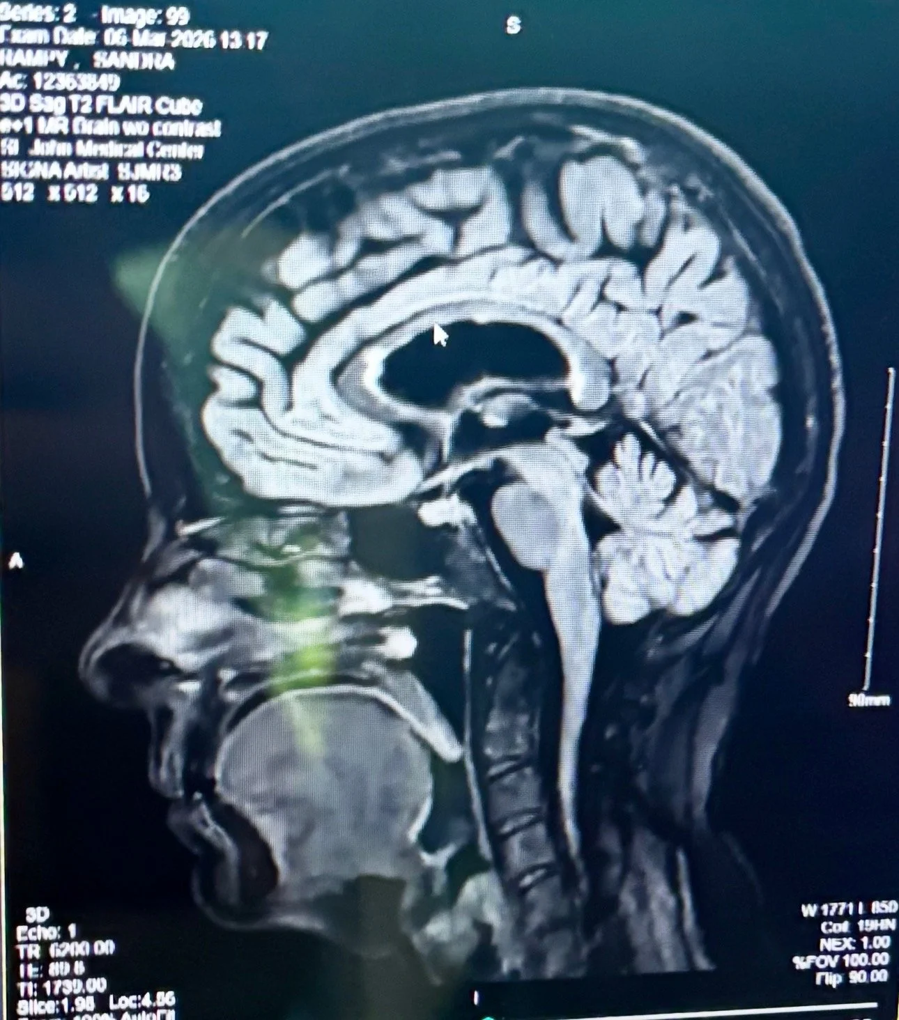
MRI scans are one of the most important tools used to help diagnose and monitor multiple sclerosis. They allow doctors to identify areas of inflammation, damage, or scarring — called lesions — within the brain and spinal cord. These images help physicians better understand disease activity, guide treatment decisions, and track changes over time.

Scientific diagram of a neuron illustrating the myelin sheath, axon, Schwann cells, dendrites, nodes of Ranvier, and nerve signal pathways. Educational resource explaining how Multiple Sclerosis damages protective nerve coverings and affects neurolog
Medical MRI scan of the human brain in sagittal view used for neurological evaluation and diagnosis of conditions such as Multiple Sclerosis. Black-and-white radiology imaging showing detailed internal brain structure

This illustration shows how myelin forms a protective coating around nerve fibers, similar to the insulation surrounding an electrical wire. Myelin helps nerve signals travel quickly and efficiently between the brain and body. In multiple sclerosis, the immune system damages this protective layer, disrupting communication within the nervous system and leading to many of the symptoms experienced in MS.